Showing posts with label X-rays. Show all posts

Sunday, 10 July 2011

MEDICAL IMAGE DATABASES

Sunday, 20 January 2008

X-RAY MACHINE DEMONSTRATION IN YEAR 1958

COIN OPERATED X-RAY MACHINE OF YEAR 1900


The glass case contains the x-ray tube and high voltage coil. stick you hand in the slot about and view your hand bones through the double eyepiece on top

WORLD'S FIRST X-RAY IMAGE


(FIRST X-RAY IMAGE OF MRS.ROENTGEN)

Wednesday, 25 July 2007

X-RAY MACHINES - PICTURES

1.)OLD X-RAY MACHINES


2.)PORTABLE X-RAY MACHINE


3.)FIXED X-RAY MACHINES


4.)DENTAL X-RAY MACHINES

i.)OLD DENTAL PANORAMIC X-RAY MACHINE


ii.)MODERN PANORAMIC DENTAL X-RAY MACHINE


iii.)WALL MOUNTED DENTAL X-RAY MACHINE


iv.)PORTABLE DENTAL X-RAY MACHINE


v.)HANDHELD DENTAL X-RAY MACHINE


5.) DUAL ENERGY X-RAY(DEXA) MACHINE / BONE DENSITOMETER


Tuesday, 24 July 2007

HANDHELD X-RAY SYSTEM - VIDEO

NOMAD is a handheld intraoral x-ray system that is cordless, battery-powered, true DC, lightweight and simple-to-use. An external backscatter shield and unique internal radiation shielding ensure operator safety that exceeds regulatory requirements. NOMAD's true portability doesn't compromise the diagnostic quality of the x-ray images. It's economical. In the office, NOMAD does the work of multiple conventional x-rays.


VIDEO OF A RADIOGRAPHER

X-RAY ATTENUATION THEORY

A photon is an X-ray when it is formed by an event involving an electron, while the photon is a gamma ray when it comes from the nucleus of an atom. In general, medical radiography is done using X-rays formed in an X-ray tube. Nuclear medicine typically involves gamma rays.

The types of electromagnetic radiation of most interest to radiography are X-ray and gamma radiation. This radiation is much more energetic than the more familiar types such as radio waves and visible light. It is this relatively high energy which makes gamma rays useful in radiography but potentially hazardous to living organisms.

The radiation is produced by X-ray tubes, high energy X-ray equipment or natural radioactive elements, such as radium and radon, and artificially produced radioactive isotopes of elements, such as cobalt-60 and iridium-192. Electromagnetic radiation consists of oscillating electric and magnetic fields, but is generally depicted as a single sinusoidal wave. While in the past radium and radon have both been used for radiography, they have fallen out of use as they are irksome radiotoxic alpha radiation emitters which are expensive; iridium-192 and cobalt-60 are far better photon sources. For further details see commonly used gamma emitting isotopes.

Such a wave is characterised by its wavelength (the distance from a point on one cycle to the corresponding point on the next cycle) or its frequency (the number of oscillations per second). In a vacuum, all electromagnetic waves travel at the same speed, the speed of light (c). The wavelength (λ, lambda) and the frequency (f) are all related by the equation:
f = c / λ

This is true for all electromagnetic radiation.

Electromagnetic radiation is known by various names, depending on its energy. The energy of these waves is related to the frequency and the wavelength by the relationship:
E = hf = h (c / λ)

Where h is a constant known as Planck's Constant.

Gamma rays are indirectly ionizing radiation. A gamma ray passes through matter until it undergoes an interaction with an atomic particle, usually an electron. During this interaction, energy is transferred from the gamma ray to the electron, which is a directly ionizing particle. As a result of this energy transfer, the electron is liberated from the atom and proceeds to ionize matter by colliding with other electrons along its path. Other times, the passing gamma ray interferes with the orbit of the electron, and slows it, releasing energy but not becoming dislodged. The atom is not ionised, and the gamma ray continues on, although at a lower energy. This energy released is usually heat or another, weaker photon, and causes biological harm as a radiation burn. The chain reaction caused by the initial dose of radiation can continue after exposure, much like a sunburn continues to damage skin even after one is out of direct sunlight.

For the range of energies commonly used in radiography, the interaction between gamma rays and electrons occurs in two ways. One effect takes place where all the gamma ray's energy is transmitted to an entire atom. The gamma ray no longer exists and an electron emerges from the atom with kinetic (motion in relation to force) energy almost equal to the gamma energy. This effect is predominant at low gamma energies and is known as the photoelectric effect. The other major effect occurs when a gamma ray interacts with an atomic electron, freeing it from the atom and imparting to it only a fraction of the gamma ray's kinetic energy. A secondary gamma ray with less energy (hence lower frequency) also emerges from the interaction. This effect predominates at higher gamma energies and is known as the Compton effect.

In both of these effects the emergent electrons lose their kinetic energy by ionizing surrounding atoms. The density of ions so generated is a measure of the energy delivered to the material by the gamma rays.

The most common means of measuring the variations in a beam of radiation is by observing its effect on a photographic film. This effect is the same as that of light, and the more intense the radiation is, the more it darkens, or exposes, the film. Other methods are in use, such as the ionizing effect measured electronically, its ability to discharge an electrostatically charged plate or to cause certain chemicals to fluoresce as in fluoroscopy.

MEDICAL RADIOGRAPHY

i.)Radiography is the use of ionising electromagnetic radiation to view objects.
ii.)The modification of objects with radiation is not radiography. In cases of modification, when the object is a human or an animal and the purpose is a cure of disease, it is radiotherapy and when the object is not living it could be food irradation or some other form of radiation processing.

Diagnostic radiography

i.)Diagnostic radiography involves the use of both ionising radiation and non-ionising radiation to create images for medical diagnoses.

ii.)The predominant test is still the X-ray (the word X-ray is often used for both the test and the actual film or digital image). X-rays are the second most commonly used medical tests, after laboratory tests. This application is known as diagnostic radiography.

iii.)Since the body is made up of various substances with differing densities, X-rays can be used to reveal the internal structure of the body on film by highlighting these differences using attenuation, or the absorption of X-ray photons by the denser substances (like calcium-rich bones). Medical diagnostic radiography is undertaken by a specially trained professional called a diagnostic radiographer or a radiologic technologist in Healthcare centers.

SUB-SPECIALITIES OF DIAGNOSTIC RADIOGRAPHY :

a.)Projection radiography
b.)Fluoroscopy (angiography, gastro-intestinal fluroscopy)
c.)Dual energy X-ray absorptiometry
d.)Computed tomography

a.)PROJECTION RADIOGRAPHY

The creation of images by exposing an object to X-rays or other high-energy forms of electromagnetic radiation and capturing the resulting remnant beam (or "shadow") as a latent image is known as "projection radiography." The "shadow" may be converted to light using a fluorescent screen, which is then captured on photographic film, it may be captured by a phosphor screen to be "read" later by a laser (CR), or it may directly activate a matrix of solid-state detectors (DR--similar to a very large version of a CCD in a digital camera). Bone and some organs (such as lungs) especially lend themselves to projection radiography. It is a relatively low-cost investigation with a high diagnostic yield.

Projection radiography uses X-rays in different amounts and strengths depending on what body part is being imaged:

i.)HARD TISSUES
Hard tissues such as bone require a relatively high energy photon source, and typically a tungsten anode is used with a high voltage (50-150 kVp) on a 3-phase or high-frequency machine to generate braking radiation. Bony tissue and metals are denser than the surrounding tissue, and thus by absorbing more of the X-ray photons they prevent the film from getting exposed as much. Wherever dense tissue absorbs or stops the X-rays, the resulting X-ray film is unexposed, and appears translucent blue, whereas the black parts of the film represent lower-density tissues such as fat, skin, and internal organs, which could not stop the X-rays. This is usually used to see bony fractures, foreign objects (such as ingested coins), and used for finding bony pathology such as osteoarthritis, infection (osteomyelitis), cancer (osteosarcoma), as well as growth studies (leg length, achondroplasia, scoliosis, etc).

ii.)SOFT TISSUES
Soft tissues are seen with the same machine as for hard tissues, but a "softer" or less-penetrating X-ray beam is used. Tissues commonly imaged include the lungs and heart shadow in a chest X-ray, the air pattern of the bowel in abdominal X-rays, the soft tissues of the neck, the orbits by a skull X-ray before an MRI to check for radiopaque foreign bodies (especially metal), and of course the soft tissue shadows in X-rays of bony injuries are looked at by the radiologist for signs of hidden trauma (for example, the famous "fat pad" sign on a fractured elbow).

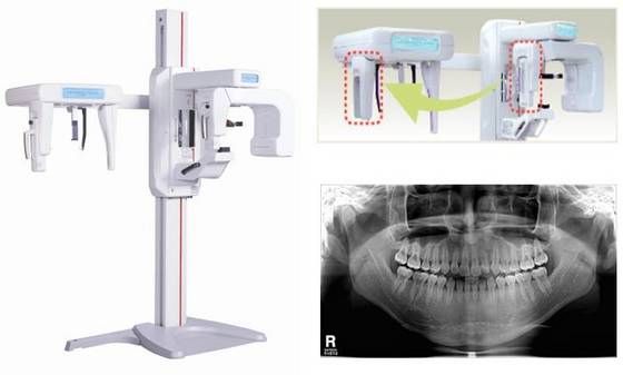
iii.)DENTAL RADIOGRAPHY
Dental radiography uses a small radiation dose with high penetration to view teeth, which are relatively dense. A dentist may examine a painful tooth and gum using X-ray equipment. The machines used are typically single-phase pulsating DC, the oldest and simplest sort. Dental technicians or the dentist may run these machines-- radiologic technologists are not required by law to be present.

iv.)MAMMOGRAPHY
Mammography is an X-ray examination of breasts and other soft tissues. This has been used mostly on women to screen for breast cancer, but is also used to view male breasts, and used in conjunction with a radiologist or a surgeon to localise suspicious tissues before a biopsy or a lumpectomy. Breast implants designed to enlarge the breasts reduce the viewing ability of mammography, and require more time for imaging as more views need to be taken. This is because the material used in the implant is very dense compared to breast tissue, and looks white (clear) on the film. The radiation used for mammography tends to be softer (has a lower photon energy) than that used for the harder tissues. Often a tube with a molybdenum anode is used with about 30 000 volts (30 kV), giving a range of X-ray energies of about 15-30 keV. Many of these photons are "characteristic radiation" of a specific energy determined by the atomic structure of the target material (Mo-K radiation).


b.)FLUOROSCOPY (angiography, gastro-intestinal fluroscopy)

Fluoroscopy is a term invented by Thomas Edison during his early X-ray studies. The name refers to the fluorescence he saw while looking at a glowing plate bombarded with X-rays.

This is a technique that provides moving projection radiographs of lower quality. Fluoroscopy is mainly performed to view movement (of tissue or a contrast agent), or to guide a medical intervention, such as angioplasty, pacemaker insertion, or joint repair/replacement. The latter are often carried out in the operating theatre, using a portable fluoroscopy machine called a C-arm. It can move around the surgery table and make digital images for the surgeon.

Angiography is the use of fluoroscopy to view the cardiovascular system. An iodine-based contrast is injected into the bloodstream and watched as it travels around. Since liquid blood and the vessels are not very dense, a contrast with high density (like the large iodine atoms) is used to view the vessels under X-ray. Angiography is used to find aneurysms, leaks, blockages (thromboses), new vessel growth, and placement of catheters and stents. Balloon angioplasty is often done with angiography.

Fluoroscopy can be used to examine the digestive system using a substance which is opaque to X-rays, (usually barium sulphate or gastrografin), which is introduced into the digestive system either by swallowing or as an enema. This is normally as part of a double contrast technique, using positive and negative contrast. Barium sulfate coats the walls of the digestive tract (positive contrast), which allows the shape of the digestive tract to be outlined as white or clear on an X-ray. Air may then be introduced (negative contrast), which looks black on the film. The barium meal is an example of a contrast agent swallowed to examine the upper digestive tract. Note that while soluble barium compounds are very toxic, the insoluble barium sulfate is non-toxic because its low solubility prevents the body from absorbing it.

A number of substances have been used as positive contrast agents: silver, bismuth, cesium, thorium, tin, zirconium, tantalum, tungsten and lanthanide compounds have been used as contrast agents. The use of thoria (thorium dioxide) as an agent was rapidly stopped as thorium causes liver cancer.

Most modern injected radiographic positive contrast media are iodine-based. Patients who suffer from allergy to shellfish may be allergic to iodine, and should consult their physician regarding pre-medication to lessen risk of allergic reaction. Iodinated contrast comes in two forms: ionic and non-ionic compounds. Non-ionic contrast is significantly more expensive than ionic (approximately three to five times the cost), however, non-ionic contrast tends to be safer for the patient, causing fewer allergic reactions and uncomfortable side effects such as hot sensations or flushing. Most imaging centers now use non-ionic contrast exclusively, finding that the benefits to patients outweigh the expense.

Negative radiographic contrast agents are air and carbon dioxide (CO2). The latter is easily absorbed by the body and causes less spasm. It can also be injected into the blood, where air absolutely cannot.

c.)Dual energy X-ray absorptiometry

DEXA, or bone densitometry, is used primarily for osteoporosis tests. It is not projection radiography, as the X-rays are emitted in 2 narrow beams that are scanned across the patient, 90 degrees from each other. Usually the hip (head of the femur), lower back (lumbar spine) or heel (calcaneum) are imaged, and the bone density (amount of calcium) is determined and given a number (a T-score). It is not used for bone imaging, as the image quality is not good enough to make an accurate diagnostic image for fractures, inflammation etc. It can also be used to measure total body fat, though this isn't common. The radiation dose received from DEXA scans is very low, much lower than projection radiography examinations.

d.)Computed tomography

Computed tomography or CT scan (previously known as CAT scan, the "A" standing for "axial") uses a high amount of ionizing radiation (in the form of X-rays) in conjunction with a computer to create images of both soft and hard tissues. These images look as though the patient was sliced like bread (thus, "tomography"-- "tomo" means "slice"). The machine looks similar to an MRI machine to many patients, but is not related. The exams are generally short, most lasting only as long as a breath-hold. Contrast agents are often used, depending on the tissues needing to be seen. Radiographers perform these examinations, sometimes in conjunction with a radiologist (for instance, when a radiologist performs a CT-guided biopsy).

Other Tests

Other tests are often found within the radiology department, due to the fact that they are all diagnostic imaging (finding disease via imagery), but do not use ionising radiation (and thus are not technically radiography). As the dangers of ionising radiation are well known, other methods of imaging were sought out and have developed over the years. These are summarised below.

Nuclear medicine

Nuclear medicine is a speciality using injected or inhaled radioactive isotopes to image the body. Instead of passing ionising radiation through the patient, the patient becomes temporarily radioactive and is placed near gamma cameras, which detect the radioactivity and store it as an image. Images made with nuclear medicine is usually physiological, not anatomical; that is, the images show function quite well, but not anatomic structure. Examples of nuclear medicine tests are PET and SPECT. Tests are most often made of the heart, thyroid gland, and cancer studies. In veterinary use, scintigraphy is commonly used for diagnosing occult bone fractures and other bony pathology in horses. Nuclear Medicine Technologists in the United States registered by the ARRT (American Registry of Radiologic Technologists) and/or certified by the NMTCB (Nuclear Medicine Technology Certification Board) perform these examinations.


Medical ultrasound

Ultrasound or sonography uses high frequency sound to create images. It is usually used to image the soft tissues of the abdomen, the pelvic area, the breasts, and the cardiovascular system, though it is also often used for guiding needles when doctors perform thoracentesis, amniocentesis, or biopsies. A specialised type of sonography is echocardiography, which specifically views the heart and surrounding major blood vessels. It has a well developed specialist training scheme, with postgraduate qualification, but is not recognised as its own profession by the Health Professions Council in the UK. Practitioners are most commonly registered diagnostic sonographers (US), radiographers (UK), doctors and midwives.

Magnetic resonance imaging (MRI)

Also known as Nuclear Magnetic Resonance (NMR) in chemistry, this is another speciality of radiography that uses non-ionising radiation, in this case magnetic fields and radio frequencies, to create images. Most lay people tell this machine apart from CT by the long exam times (several minutes to an hour), the claustrophobic tunnel, and the loud pounding noise created by the growing and collapsing of the magnetic fields. Specially trained radiographers carry out these examinations. Post-graduate training is necessary to practice in this field for several reasons. The physics of MRI are slightly different and quite a bit more complex than in traditional (plain-film/screen, CR, DR) or even CT imaging modalities. Sectional soft tissue details when using MR have a different appearance than those in CT, requiring additional training to allow the technologist to become familiar with the appearances of both normal and pathologic anatomy which may require adjustment of technique. Finally the risk of serious injury to the patient far surpasses any such risk present in other imaging modalities. Magnetic field strength in medical MRI scanners range from 1.5 to 3 teslas on average. These fields would easily turn any ferromagnetic object into a projectile (this includes some necklaces, earrings, surgical pins, etc.) thus as the person actually conducting the examination, the technologist must ensure and be familiar with a full medical history to rule out implanted ferromagnetic objects, as well as (of course) ensuring no such objects (implanted or otherwise) enter the room with the patient. Although this may not seem like a difficult task, this risk of serious injury or death is very real, making this a major professional responsibility.

Radiotherapy and therapeutic radiography

As part of the planning and execution of radiotherapy radiography is often used as part of the treatment of cancer in oncology departments, with 6 out of 10 patients treated with radiotherapy for cancer cured. This involves application of a prescribed dose of ionising radiation to specific targeted tissue, whilst limiting damage to the surrounding healthy area.

This treatment is prescribed by an oncologist and is conducted by radiotherapists, who are a group of professions working together, including medical physicists, therapy radiographers, and technicians.

X-RAY MACHINES


1.)An X-ray machine utilizes electromagnetic radiation to produce an image of an object, usually with the purpose of visualizing something located below the object's surface.
2.)The machine is made up of an X-ray source or X-ray tube, an x-ray detection system, and positioning hardware to align these two components with the object to be imaged.

BASICS OF X-RAY MACHINE


An X-ray imaging system consists of a X-ray source or generator, and an image detection system which can either be comprised of film (analog technology) or a digital capture system.

X-ray Sources

i.)In the typical X-ray source of less than 450kV, X-ray photons are produced by an electron beam striking a target. The electrons that make up the stream beam are emitted from a heated cathode filament. The electrons are then focused and accelerated towards an anode target. The point where the electron beam strikes the target is called the focal spot. Most of the kinetic energy contained in the electron beam is converted to heat. But a small percentage is converted into X-ray. At the focal spot, X-ray photons are emitted in all directions including towards a small window in the X-ray tube. This window allows the X-ray to exit the tube with little attenuation while maintaining a vacuum seal required for the X-ray tube operation.

ii.)X-ray machines work by applying controlled voltage, current, and time to the X-ray tube, which results in a beam of X-rays. The beam is projected on matter. Some of the X-ray beam will pass through the object, while some is reflected. The resulting pattern of the radiation is then ultimately detected by a detection medium including rare earth screens (which surround photographic film), semiconductor detectors, or X-ray image intensifiers.

X-Ray Detection Systems

In healthcare applications in particular, the x-ray detection system rarely consists of the detection medium. For example, a typical stationary radiographic x-ray machine also includes an ion chamber and grid. The ion chamber is basically a hollow plate located between the detection medium and the object being imaged. It determines the level of exposure by measuring the amount of x-rays that have passed through the electrically charged, gas-filled gap inside the plate. This allows for minimization of patient radiation exposure by both ensuring that an image is not underdeveloped to the point the exam needs to be repeated and ensuring that more radiation than needed is not applied. The grid is usually located between the ion chamber and object and consists of several lead slats stacked next to each other (resembling open window blinds). In this manner, the grid allows straight x-rays to pass through to the detection medium but absorbs reflected x-rays. This improves image quality by preventing reflected (non-diagnostic) x-rays from reaching the detection medium allowing for lower exam doses overall.

Images taken with such devices are known as X-ray photographs or radiographs.

USES

a.) MEDICAL USES
There are two basic areas in which Health Care uses X-radiation; Radiography, and Fluoroscopy.

i.)Radiography is used for fast, highly penetrating images. Usually it's used on areas with a high bone content. Some forms of radiography uses are Panoramic X-rays, Radiography, Mammography, Tomography, and Radiotherapy.

ii.)Fluoroscopy is used in cases where real-time visualization is necessary. You may have seen a type of fluorography at the airport. Some of the uses of Fluorography are Angiography, barium enemas, barium swallows, biopsies, and hip replacement.

iii.)X-rays are highly penetrating, ionizing radiation, and X-ray machines are used in radiology to take pictures of bones and teeth. This is because bones absorb the radiation more than the less-dense soft tissue. X-rays from a source pass through the body and onto a photographic cassette. Areas where radiation is absorbed show up as lighter shades of gray (closer to white). This can be used to diagnose broken or fractured bones.

iv.)Imaging of the digestive tract is done with the help of a radiocontrast agent such as barium sulfate, which is opaque to X-rays.

b.) SECURITY SYSTEMS


X-ray machines are used to screen objects non-invasively. Luggage at airports is examined for possible bombs and weapons. These machines are very low dose and safe to be around.The largest manufacturer of X-Ray inspection systems is Smiths Heimann GmbH located in Wiesbaden, Germany.

DETECTORS OF X-RAYS

1.)Photographic Plate

i.)The detection of X-rays is based on various methods. The most commonly known method are a photographic plate, X-ray film in a cassette, and rare earth screens.

ii.)A photographic plate or film is used in hospitals to produce images of the internal organs and bones of a patient. Since photographic plates are not generally sensitive to X-rays, phosphorescent screens are usually placed in contact with the emulsion of the plate or film. The X-rays strike the phosphor screen, which emits visible light, which exposes the film. The emulsion still needs to be heavily doped with silver compounds and can be coated on both sides of the film or plate.

iii.)The part of the patient to be X-rayed is placed between the X-ray source and the photographic receptor to produce what is a shadow of all the internal structure of that particular part of the body being X-rayed. The X-rays are blocked by dense tissues such as bone and pass through soft tissues. Those areas where the X-rays strike the photographic receptor turn black when it is developed. So where the X-rays pass through "soft" parts of the body such as organs, muscle, and skin, the plate or film turns black.

iv.)Contrast compounds containing barium or iodine, which are radiopaque, can be injected in the artery of a particular organ, or given intravenously. The contrast compounds essentially block the X-rays and hence the circulation of the organ can be more readily seen. Many years ago thorium was used as a contrast medium (Thorotrast) - this caused many people to be injured or even die from the effects of the radiation from the thorium.


2.)Photostimulable Phosphors (PSPs)

An increasingly common method of detecting X-rays is the use of Photostimulable Luminescence (PSL), pioneered by Fuji in the 1980s.

In modern hospitals a PSP plate is used in place of the photographic plate. After the plate is X-rayed, excited electrons in the phosphor material remain 'trapped' in 'colour centres' in the crystal lattice until stimulated by a laser beam passed over the plate surface. The light given off during laser stimulation is collected by a photomultiplier tube and the resulting signal is converted into a digital image by computer technology, which gives this process its common name, computed radiography (also referred to as digital radiography). The PSP plate can be used over and over again.


3.)Geiger counter

i.)Initially, most common detection methods were based on the ionization of gases, as in the Geiger-Müller counter: a sealed volume, usually a cylinder, with a polymer or thin metal window contains a gas, and a wire, and a high voltage is applied between the cylinder (cathode) and the wire (anode). When an X-ray photon enters the cylinder, it ionizes the gas and forms ions and electrons. Electrons accelerate toward the anode, in the process causing further ionization along their trajectory. This process, known as an avalanche, is detected as a sudden flow of current, called a "count" or "event".

ii.)Ultimately, the electrons form a virtual cathode around the anode wire drastically reducing the electric field in the outer portions of the tube. This halts the collisional ionizations and limits further growth of avalanches. As a result, all "counts" on a Geiger counter are the same size and it can give no indication as to the particle energy of the radiation, unlike the proportional counter. The intensity of the radiation is measurable by the Geiger counter as the counting-rate of the system.

iii.)In order to gain energy spectrum information a diffracting crystal may be used to first separate the different photons, the method is called wavelength dispersive X-ray spectroscopy (WDX or WDS).

iv.)Position-sensitive detectors are often used in conjunction with dispersive elements. Other detection equipment may be used which are inherently energy-resolving, such as the aforementioned proportional counters. In either case, use of suitable pulse-processing (MCA) equipment allows digital spectra to be created for later analysis.

v.)For many applications, counters are not sealed but are constantly fed with purified gas (thus reducing problems of contamination or gas aging). These are called "flow counter".


4.)Scintillators

Some materials such as sodium iodide (NaI) can "convert" an X-ray photon to a visible photon; an electronic detector can be built by adding a photomultiplier. These detectors are called "scintillators", filmscreens or "scintillation counters". The main advantage of using these is that an adequate image can be obtained while subjecting the patient to a much lower dose of X-rays.


5.) Image Intensification

X-rays are also used in "real-time" procedures such as angiography or contrast studies of the hollow organs (e.g. barium enema of the small or large intestine) using fluoroscopy acquired using an X-ray image intensifier. Angioplasty, medical interventions of the arterial system, rely heavily on X-ray-sensitive contrast to identify potentially treatable lesion.


6.)Direct Semiconductor Detectors

Since the 1970s, new semiconductor detectors have been developed (silicon or germanium doped with lithium, Si(Li) or Ge(Li)).

i.)X-ray photons are converted to electron-hole pairs in the semiconductor and are collected to detect the X-rays. When the temperature is low enough (the detector is cooled by Peltier effect or best by liquid nitrogen), it is possible to directly determine the X-ray energy spectrum; this method is called energy dispersive X-ray spectroscopy (EDX or EDS); it is often used in small X-ray fluorescence spectrometers. These detectors are sometimes called "solid detectors". Cadmium telluride (CdTe) and its alloy with zinc, cadmium zinc telluride detectors have an increased sensitivity, which allows lower doses of X-rays to be used.

ii.)Currently amorphous selenium is used in commercial large area flat panel X-ray detectors for chest radiography and mammography.

Note:
A standard semiconductor diode, such as a 1N4007, will produce a small amount of current when placed in an X-ray beam. A test device once used by Medical Imaging Service personnel was a small project box that contained several diodes of this type in series, which could be connected to an oscilloscope as a quick diagnostic.

iii.)Silicon drift detectors (SDDs), produced by conventional semiconductor fabrication, now provide a cost-effective and high resolving radiation measurement. They replace conventional X-ray detectors, such as Si(Li)s, as they do not need to be cooled with liquid nitrogen.


7.)Scintillator + semiconductor detectors (indirect detection)

i.)With the advent of large semiconductor array detectors it has become possible to design detector systems using a scintillator screen to convert from X-rays to visible light which is then converted to electrical signals in an array detector. Indirect Flat Panel Detectors (FPDs) are in widespread use today in medical, dental, veterinary and industrial applications. A common form of these detectors is based on amorphous silicon TFT/photodiode arrays.

ii.)The array technology is a variant on the amorphous silicon TFT arrays used in many flat panel displays, like the ones in computer laptops. The array consists of a sheet of glass covered with a thin layer of silicon that is in an amorphous or disordered state. At a microscopic scale, the silicon has been imprinted with millions of transistors arranged in a highly ordered array, like the grid on a sheet of graph paper. Each of these thin film transistors (TFTs) are attached to a light-absorbing photodiode making up an individual pixel (picture element).

ii.)Photons striking the photodiode are converted into two carriers of electrical charge, called electron-hole pairs. Since the number of charge carriers produced will vary with the intensity of incoming light photons, an electrical pattern is created that can be swiftly converted to a voltage and then a digital signal, which is interpreted by a computer to produce a digital image.

iii.)Although silicon has outstanding electronic properties, it is not a particularly good absorber of X-ray photons. For this reason, X-rays first impinge upon scintillators made from eg. gadolinium oxysulfide or cesium iodide. The scintillator absorbs the X-rays and converts them into visible light photons that then pass onto the photodiode array.

8.)Visibility to the human eye

While generally considered invisible to the human eye, in special circumstances X-rays can be visible. Brandes, in an experiment a short time after Röntgen's landmark 1895 paper, reported after dark adaptation and placing his eye close to an X-ray tube, seeing a faint "blue-gray" glow which seemed to originate within the eye itself. Upon hearing this, Röntgen reviewed his record books and found he too had seen the effect. When placing an X-ray tube on the opposite side of a wooden door Röntgen had noted the same blue glow, seeming to emanate from the eye itself, but thought his observations to be spurious because he only saw the effect when he used one type of tube. Later he realized that the tube which had created the effect was the only one powerful enough to make the glow plainly visible and the experiment was thereafter readily repeatable. The knowledge that X-rays are actually faintly visible to the dark-adapted naked eye has largely been forgotten today; this is probably due to the desire not to repeat what would now be seen as a recklessly dangerous and harmful experiment with ionizing radiation. It is not known what exact mechanism in the eye produces the visibility: it could be due to conventional detection (excitation of rhodopsin molecules in the retina), direct excitation of retinal nerve cells, or secondary detection via, for instance, X-ray induction of phosphorescence in the eyeball with conventional retinal detection of the secondarily produced visible light.

If the intensity of a X-ray beam is high enough, the ionization of the air will make the beam visible with a white glow.

USES OF X-RAYS

MEDICAL USES
Since it is discovered that X-rays can identify bony structures, X-rays have been developed for their use in medical imaging. Radiology is a specialized field of medicine. Radiographers employ radiography and other techniques for diagnostic imaging. This is probably the most common use of X-ray technology.

1.)SKELETAL SYSTEM
X-rays are especially useful in the detection of pathology of the skeletal system, but are also useful for detecting some disease processes in soft tissue.

2.)CHEST X-RAY
Some notable examples are the very common chest X-ray, which can be used to identify lung diseases such as pneumonia, lung cancer or pulmonary edema

3.)ABDOMINAL X-RAY
the abdominal X-ray, which can detect ileus (blockage of the intestine), free air (from visceral perforations) and free fluid (in ascites).

4.)DETECTING STONES IN BODY
In some cases, the use of X-rays is debatable, such as gallstones (which are rarely radiopaque) or kidney stones (which are often visible, but not always).

5.)Also, traditional plain X-rays pose very little use in the imaging of soft tissues such as the brain or muscle. Imaging alternatives for soft tissues are computed axial tomography (CAT or CT scanning), magnetic resonance imaging (MRI) or ultrasound.

6.)RADIOTHERAPY
Radiotherapy, a curative medical intervention, now used almost exclusively for cancer, employs higher energies of radiation.

OTHER USES OF X-RAYS

1.)X-ray crystallography
X-ray crystallography in which the pattern produced by the diffraction of X-rays through the closely spaced lattice of atoms in a crystal is recorded and then analyzed to reveal the nature of that lattice.

2.)X-ray astronomy
X-ray astronomy, which is an observational branch of astronomy, which deals with the study of X-ray emission from celestial objects.

3.)X-ray microscopy
X-ray microscopic analysis, which uses electromagnetic radiation in the soft X-ray band to produce images of very small objects.

4.)X-ray fluorescopy
X-ray fluorescence, a technique in which X-rays are generated within a specimen and detected. The outgoing energy of the X-ray can be used to identify the composition of the sample.

5.)Paintings are often X-rayed to reveal the underdrawing and pentimenti or alterations in the course of painting, or by later restorers. Many pigments such as lead white show well in X-ray photographs

DIGITAL ANGIOGRAPHY

To create a blood or artery X-ray, also called digital angiography, iodine is injected into the veins and a digitized image is created. Then, a second image is established of only the parts of the X-rayed section without iodine. The first image is subtracted then a final image is produced containing both the first and second images together. Lastly, the results are printed. The doctor or surgeon then compares the results of the angiography to a perfect angiography structure to see if there are any malfunctions.

X-RAYS

1.) X-rays (or Röntgen rays) are a form of electromagnetic radiation with a wavelength in the range of 10 to 0.01 nanometers, corresponding to frequencies in the range 30 to 30 000 PHz (1 PHz = 1015 Hertz).
2.)X-rays are primarily used for diagnostic radiography and crystallography. X-rays are a form of ionizing radiation and as such can be dangerous.

UNITS OF MEASURE


1.)The unit of measure for an X-ray is called a rem, "r".
2.)A rem is a relatively large amount, and therefore exposure to X-rays for medical use is often measured in mrems (or millirems).

PHYSICS OF X-RAYS


1.)i.)X-rays are a type of electromagnetic radiation with wavelengths of around 10-10 meters.
ii.)When medical X-rays are being produced, a thin metallic sheet is placed between the emitter and the target, effectively filtering out the lower energy (soft) X-rays. This is often placed close to the window of the X-ray tube. The resultant X-ray is said to be hard.
iii.)Soft X-rays overlap the range of extreme ultraviolet. The frequency of hard X-rays is higher than that of soft X-rays, and the wavelength is shorter.
iv.)Hard X-rays overlap the range of "long"-wavelength (lower energy) gamma rays, however the distinction between the two terms depends on the source of the radiation, not its wavelength;
v.)X-ray photons are generated by energetic electron processes, gamma rays by transitions within atomic nuclei.

2.)PRODUCTION OF X-RAYS
i.)The basic production of X-rays is by accelerating electrons in order to collide with a metal target. (In medical applications, this is usually tungsten or a more crack resistant alloy of rhenium (5%) and tungsten (95%), but sometimes molybdenum for more specialised applications, such as when soft X-rays are needed as in mammography. In crystallography, a copper target is most common, with cobalt often being used when fluorescence from iron content in the sample might otherwise present a problem.)

ii.)Here the electrons suddenly decelerate upon colliding with the metal target and if enough energy is contained within the electron it is able to knock out an electron from the inner shell of the metal atom and as a result electrons from higher energy levels then fill up the vacancy and X-ray photons are emitted. This process is extremely inefficient (~0.1%) and thus to produce reasonable flux of X-rays plenty of energy has to be wasted into heat which has to be removed.

iii.)The spectral lines generated depends on the target (anode) element used and thus are called characteristic lines. Usually these are transitions from upper shells into K shell (called K lines), into L shell (called L lines) and so on. There is also a continuum Bremsstrahlung radiation given off by the electrons as they are scattered by the strong electric field near the high-Z (proton number) nuclei.

3.)X-rays can detect cancer, cysts, and tumors. Due to their short wavelength X-rays act more like a particle than a wave.

Nowadays, for many (non medical) applications, X-ray production is achieved by synchrotrons .

DIGITAL ANGIOGRAPHY

To create a blood or artery X-ray, also called digital angiography, iodine is injected into the veins and a digitized image is created. Then, a second image is established of only the parts of the X-rayed section without iodine. The first image is subtracted then a final image is produced containing both the first and second images together. Lastly, the results are printed. The doctor or surgeon then compares the results of the angiography to a perfect angiography structure to see if there are any malfunctions.

To take an X-ray of the bones, no iodization is required. Short X-ray pulses are shot through a body at first. Next, the bones absorb the most waves because they are more dense and contain Ca which absorbs stronger than C,O,N atoms of soft tissue (due to more electrons in Ca atom). The X-ray film see the bones through the X-ray.

BIOMEDICAL BOOKS

Join me on Facebook Follow me on Twitter Subscribe to RSS