Showing posts with label Pictures. Show all posts

Friday, 26 September 2008

HIP IMPLANT


The above picture shows a hip implant including titanium stems, necks, sleeves, shells, and glenoid heads

PARTS OF A HIP IMPLANT


a hip prosthesis has three parts:

1.)The stem, usually made from metal
2.)The ball or head, made of ceramic or metal
3.)The shell and accompanying liner, with the shell made of metal and the liner made of a plastic called polyethylene. This liner may also be made of ceramic or metal.

Materials for implants in hip replacements include :

1.)Ceramic-on-Polyethylene (referring to a ceramic head rotating on a polyethylene liner)
2.)Metal-on-Polyethylene
3.)Ceramic-on-Ceramic
4.)Metal-on-Metal

Thursday, 9 August 2007

ELECTROCARDIOGRAM

1.)An electrocardiogram (ECG or EKG, abbreviated from the German Elektrokardiogram) is a graphic produced by an electrocardiograph, which records the electrical activity of the heart over time.

ECG WAVES

2.)Analysis of the various waves and normal vectors of depolarization and repolarization yields important diagnostic information.

PATIENT WITH ECG RECORDER

 ECG INTERPRETATION

ADVANTAGES

1.)It is the gold standard for the diagnosis of cardiac arrhythmias.

CARDIAC ARRHYTHMIAS1

CARDIA ARRHYTHMIAS2

2.)It guides therapy and risk stratification for patients with suspected acute myocardial infarction.

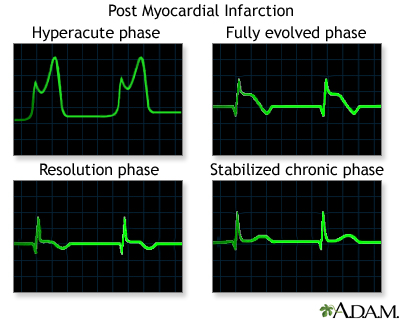
MYOCARDIAL INFARCTION ECG WAVES

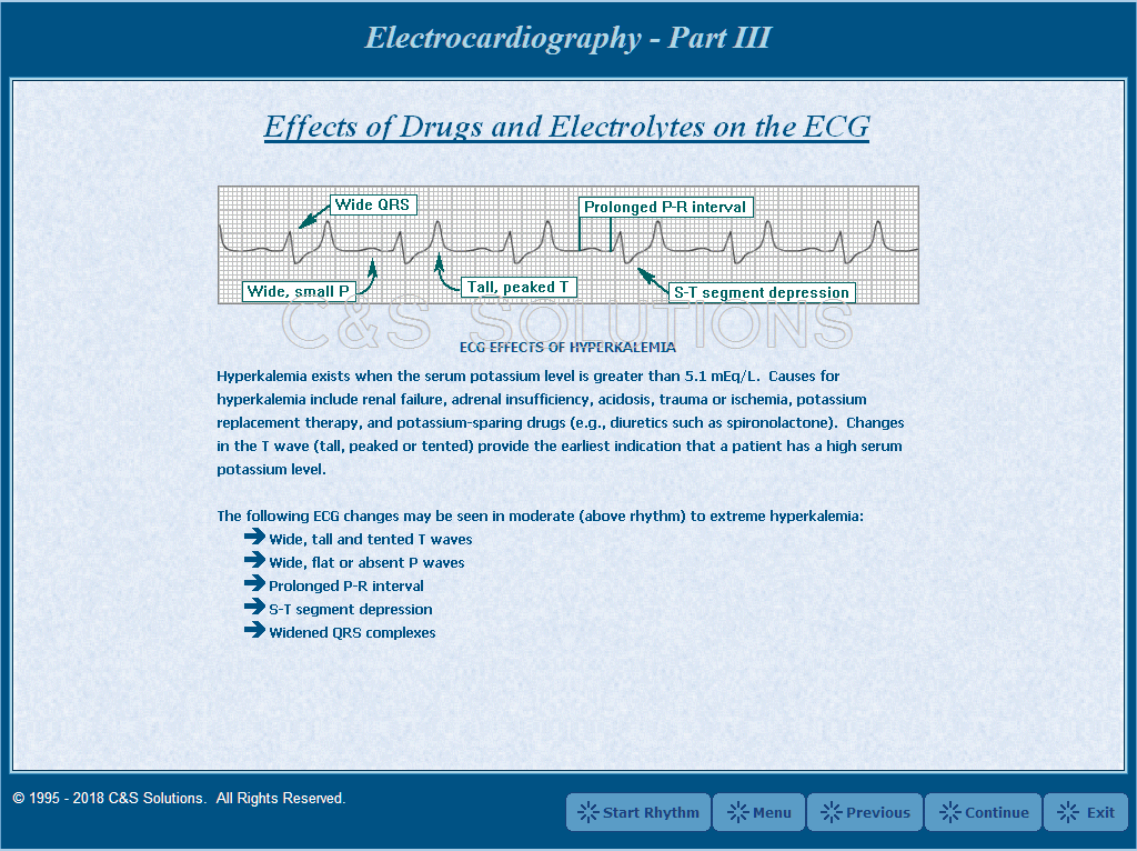
3.)It helps detect electrolyte disturbances (e.g. hyperkalemia and hypokalemia).

HYPERKALEMIA ECG

 HYPOKALEMIA & HYPERKALEMIA

4.)It allows for the detection of conduction abnormalities (e.g. right and left bundle branch block).

RIGHT BUNDLE BRANCH BLOCK

LEFT BUNDLE BRANCH BLOCK

5.)It is used as a screening tool for ischemic heart disease during a cardiac stress test.

ISCHEMIC ECG WAVE

6.)It is occasionally helpful with non-cardiac diseases (e.g. pulmonary embolism or hypothermia).

HYPOTHERMIA

PULMONARY EMBOLISM
7.)The electrocardiogram does not directly assess the contractility of the heart. However, it can give a rough indication of increased or decreased contractility.

CONTRACTION OF HEART

Sunday, 5 August 2007

EINTHOVEN ECG TRIANGLE

12 LEAD PLACEMENT EINTHOVEN ECG TRIANGLE

12 LEAD ECG PLACEMENT & INDIVIDUAL WAVEFORM

3 LEAD PLACEMENT EINTHOVEN TRIANGLE

Wednesday, 25 July 2007

X-RAY MACHINES - PICTURES

1.)OLD X-RAY MACHINES


2.)PORTABLE X-RAY MACHINE


3.)FIXED X-RAY MACHINES


4.)DENTAL X-RAY MACHINES

i.)OLD DENTAL PANORAMIC X-RAY MACHINE


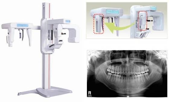
ii.)MODERN PANORAMIC DENTAL X-RAY MACHINE


iii.)WALL MOUNTED DENTAL X-RAY MACHINE


iv.)PORTABLE DENTAL X-RAY MACHINE


v.)HANDHELD DENTAL X-RAY MACHINE


5.) DUAL ENERGY X-RAY(DEXA) MACHINE / BONE DENSITOMETER


Monday, 23 July 2007

FLUOROSCOPY


Fluoroscopy is an imaging technique commonly used by physicians to obtain real-time images of the internal structures of a patient through the use of a fluoroscope. In its simplest form, a fluoroscope consists of an x-ray source and fluorescent screen between which a patient is placed. However, modern fluoroscopes couple the screen to an x-ray image intensifier and CCD video camera allowing the images to be played and recorded on a monitor. The use of x-rays, a form of ionizing radiation, requires that the potential risks from a procedure be carefully balanced with the benefits of the procedure to the patient. While physicians always try to use low dose rates during fluoroscopy procedures, the length of a typical procedure often results in a relatively high absorbed dose to the patient. Recent advances include the digitization of the images captured and flat-panel detector systems which reduce the radiation dose to the patient still further.

Fluoroscope design


The first fluoroscopes consisted of an x-ray source and fluorescent screen between which the patient would be placed. As the x rays pass through the patient, they are attenuated by varying amounts as they interact with the different internal structures of the body, casting a shadow of the structures on the fluorescent screen. Images on the screen are produced as the unattenuated x rays interact with atoms in the screen through the photoelectric effect, giving their energy to the electrons. While much of the energy given to the electrons is dissipated as heat, a fraction of it is given off as visible light, producing the images. Early radiologists would adapt their eyes to view the dim fluoroscopic images by sitting in darkened rooms, or by wearing red adaptation goggles.

X-ray Image Intensifiers

The invention of X-ray image intensifiers in the 1950s allowed the image on the screen to be visible under normal lighting conditions, as well as providing the option of recording the images with a conventional camera. Subsequent improvements included the coupling of, at first, video cameras and, later, CCD cameras to permit recording of moving images and electronic storage of still images.

Modern image intensifiers no longer use a separate fluorescent screen. Instead, a cesium iodide phosphor is deposited directly on the photocathode of the intensifier tube. On a typical general purpose system, the output image is approximately 105 times brighter than the input image. This brightness gain comprises a flux gain (amplification of photon number) and minification gain (concentration of photons from a large input screen onto a small output screen) each of approximately 100. This level of gain is sufficient that quantum noise, due to the limited number of x-ray photons, is a significant factor limiting image quality.

Image intensifiers are available with input diameters of up to 45 cm, and a resolution of approximately 2-3 line pairs mm-1.

Flat-panel detectors

The introduction of flat-panel detectors allows for the replacement of the image intensifier in fluoroscope design. Flat panel detectors offer increased sensitivity to X-rays, and therefore have the potential to reduce patient radiation dose. Temporal resolution is also improved over image intensifiers, reducing motion blurring. Contrast ratio is also improved over image intensifiers: flat-panel detectors are linear over a very wide latitude, whereas image intensifiers have a maximum contrast ratio of about 35:1. Spatial resolution is approximately equal, although an image intensifier operating in 'magnification' mode may be slightly better than a flat panel.

Flat panel detectors are considerably more expensive to purchase and repair than image intensifiers, so their uptake is primarily in specialties that require high-speed imaging, e.g., vascular imaging and cardiac catheterization.

Imaging concerns

In addition to spatial blurring factors that plague all x-ray imaging devices, caused by such things as Lubberts effect, K-fluorescence reabsorption and electron range, fluoroscopic systems also experience temporal blurring due to system lag. This temporal blurring has the effect of averaging frames together. While this helps reduce noise in images with stationary objects, it creates motion blurring for moving objects. Temporal blurring also complicates measurements of system performance for fluoroscopic systems.

Common procedures using fluoroscopy

1.)Investigations of the gastrointestinal tract, including barium enemas, barium meals and barium swallows, and enteroclysis.
2.)Orthopaedic surgery to guide fracture reduction and the placement of metalwork.
3.)Angiography of the leg, heart and cerebral vessels.
4.)Placement of a PICC (peripherally inserted central catheter)
5.)Placement of a weighted feeding tube (e.g. Dobhoff) into the duodenum after previous attempts without fluoroscopy have failed.
6.)Urological surgery – particularly in retrograde pyelography.
7.)Implantation of cardiac rhythm management devices (pacemakers, implantable cardioverter defibrillators and cardiac resynchronization devices)

Another common procedure is the modified barium swallow study during which barium-impregnated liquids and solids are ingested by the patient. A radiologist records and, with a speech pathologist, interprets the resulting images to diagnose oral and pharyngeal swallowing dysfunction. Modified barium swallow studies are also used in studying normal swallow function.

Risks

Because fluoroscopy involves the use of x rays, a form of ionizing radiation, all fluoroscopic procedures pose a potential health risk to the patient. Radiation doses to the patient depend greatly on the size of the patient as well as length of the procedure, with typical skin dose rates quoted as 20-50 mGy/min. Exposure times vary depending on the procedure being performed, but procedure times up to 75 minutes have been documented. Because of the long length of some procedures, in addition to standard cancer-inducing stochastic radiation effects, deterministic radiation effects have also been observed ranging from mild erythema, equivalent of a sun burn, to more serious burns.

A study has been performed by the Food and Drug Administration (FDA) entitled Radiation-induced Skin Injuries from Fluoroscopy with an additional publication to minimize further fluoroscopy-induced injuries, Public Health Advisory on Avoidance of Serious X-Ray-Induced skin Injuries to Patients During Fluoroscopically-Guided Procedures.

While deterministic radiation effects are a possibility, radiation burns are not typical of standard fluoroscopic procedures. Most procedures sufficiently long in length to produce radiation burns are part of necessary life-saving operations.

NOTES:

1.)ABSORBED DOSE

Absorbed dose (also known as Total Ionizing Dose, TID) is a measure of the energy deposited in a medium by ionizing radiation. It is equal to the energy deposited per unit mass of medium, and so has the unit J/kg, which is given the special name Gray (Gy).

Note that the absorbed dose is not a good indicator of the likely biological effect. 1 Gy of alpha radiation would be much more biologically damaging than 1 Gy of photon radiation for example. Appropriate weighting factors can be applied reflecting the different relative biological effects to find the equivalent dose.

The risk of stochastic effects due to radiation exposure can be quantified using the effective dose, which is a weighted average of the equivalent dose to each organ depending upon its radiosensitivity.

When ionising radiation is used to treat cancer, the doctor will usually prescribe the radiotherapy treatment in Gy. When risk from ionising radiation is being discussed, a related unit, the Sievert is used.

2.)Specific absorption rate
Specific Absorption Rate (SAR) is a measure of the rate at which radio frequency (RF) energy is absorbed by the body when exposed to radio-frequency electromagnetic field. The most common use is in relation to cellular telephones. In the United States, the Federal Communications Commission (FCC) has adopted limits for safe exposure to RF energy produced by mobile devices and requires that phones sold in the U.S. have a SAR level at or below 1.6 watts per kilogram (W/kg) taken over a volume of 1 gram of tissue. In the EU the corresponding limit is 2 W/kg (averaged over ten grams of tissue).

Tuesday, 10 July 2007

ENDOSCOPE
ENDOSCOPY
Endoscopy means looking inside and typically refers to looking inside the human body for medical reasons using an instrument called an endoscope. Endoscopy can also refer to using a borescope in engineering and technical situations where direct line-of-sight observation is not feasible.

DETAILS
1.)Endoscopy is a minimally invasive diagnostic medical procedure used to assess the interior surfaces of an organ by inserting a tube into the body. The instrument may have a rigid or flexible tube and not only provide an image for visual inspection and photography, but also enable taking biopsies and retrieval of foreign objects. Endoscopy is the vehicle for minimally invasive surgery.

2.)Many endoscopic procedures are considered to be relatively painless and, at worst, associated with mild discomfort. Most patients tolerate the procedure with only topical anaesthesia of the oropharynx using lignocaine spray.Complications are rare (only 5% of all operations)but can include perforation of the organ under inspection with the endoscope or biopsy instrument. If that occurs open surgery may be required to repair the injury.


Components
An endoscope can consist of

1.) a rigid or flexible tube
2.) a light delivery system to illuminate the organ or object under inspection. The light source is normally outside the body and the light is typically directed via an optical fiber system
3.) a lens system transmitting the image to the viewer from the fiberscope
an additional channel to allow entry of medical instruments or manipulators

Uses
Endoscopy can involve

1.) The gastrointestinal tract (GI tract):
esophagus, stomach and duodenum (esophagogastroduodenoscopy)
2.) Small intestine
colon (colonoscopy,proctosigmoidoscopy)
3.) Bile duct
endoscopic retrograde cholangiopancreatography (ERCP), duodenoscope-assisted cholangiopancreatoscopy, intraoperative cholangioscopy
4.) The respiratory tract
The nose (rhinoscopy)
The lower respiratory tract (bronchoscopy)
5.) The urinary tract (cystoscopy)
6.) The female reproductive system
The cervix (colposcopy)
The uterus (hysteroscopy)
The Fallopian tubes (Falloscopy)
7.) Normally closed body cavities (through a small incision):
The abdominal or pelvic cavity (laparoscopy)
The interior of a joint (arthroscopy)
8.) Organs of the chest (thoracoscopy and mediastinoscopy)
9.) During pregnancy
The amnion (amnioscopy)
The fetus (fetoscopy)
10.)Plastic Surgery
11.)Panendoscopy (or triple endoscopy)
Combines laryngoscopy, esophagoscopy, and bronchoscopy
12.)Non-medical uses for endoscopy
The planning and architectural community have found the endoscope useful for pre-visualization of scale models of proposed buildings and cities (architectural endoscopy)
Internal inspection of complex technical systems (borescope)
Endoscopes are also a tool helpful in the examination of improvised explosive devices by bomb disposal personel
FBI uses endoscopes to shove under doors to spy on people.

Risks
1.)Infection
2.)Punctured organs
3.)Allergic reactions due to Contrast agents or dyes (such as those used in a CT scan)
4.)Over-sedation

After The Endoscopy
After the procedure the patient will be observed and monitored by a qualified individual in the endoscopy or a recovery area until a significant portion of the medication has worn off. Occasionally a patient is left with a mild sore throat, which promptly responds to saline gargles, or a feeling of distention from the insufflated air that was used during the procedure. Both problems are mild and fleeting. When fully recovered, the patient will be instructed when to resume his/her usual diet (probably within a few hours) and will be allowed to be taken home. Because of the use of sedation, most facilities mandate that the patient is taken home by another person and not to drive on his/her own or handle machinery for the remainder of the day.


Recent developments
With the application of robotic systems, telesurgery was introduced as the surgeon could operate from a site physically removed from the patient. The first transatlantic surgery has been called the Lindbergh Operation.

Sunday, 8 July 2007

CAPSULE ENDOSCOPE


1.) Wireless capsule endoscopy, also known as the capsule camera or video pill or Miniature ingestible Capsule is a camera with the size and shape of pill used to visualize the gastrointestinal tract.

2.) This device is being promoted as an alternative to an endoscopy and has become a valuable tool to gastroenterologists all over the world with sales over 135 million dollars per year with over half million Capsules already sold. The camera assists in detecting cancer, ulcers and other types of internal medical ailments.

3.) Capsule Endoscopy is a revolutionary new technology that allows our physicians to see the middle part of your intestinal tract – the small intestine – where no scope can currently go. Our patients can now swallow a wireless video camera about the size of a large vitamin, and then go normally about their day while the capsule records images throughout the digestive tract. This new tool is especially helpful in finding the source of unexplained intestinal bleeding and for detecting Crohn’s Disease.


(Endoscopic capsule end-on, showing six LEDs and camera lens.)

4.) The Wireless Capsule endoscope can be safely ingested into human Gastrointestinal tracts and the camera using CCD or CMOS technology with long battery life takes picture and transmits them to an outside device for viewing. Battery life is key as the Capsule takes longer than 8 hours to reach Colon.

5.) At the present time, the capsule camera is primarily used to visualize the small intestine. Whereas the upper gastrointestinal tract (esophagus, stomach, and duodenum) and the colon (large intestine) can be very adequately visualized with scopes (cameras placed at the ends of thin flexible tubes), the small intestine is very long (average 20-25 feet) and very convoluted. The capsule camera travels through the length of the small intestine in about 4 hours, and wirelessly transmits two images every second to a receiver carried by the patient. The images are of very good quality, comparable to those from scopes. The test carries a high sensitivity and specificity for detecting lesions. The main uses today are for detecting the cause of gastrointestinal bleeding, and for inflammatory bowel disease, such as Crohn's disease.

5.) The Endoscopy Capsule is going to be used for many more active procedures and functions like the Current Endoscopes very soon .


RF SYSTEM LAB'S NEXT GENERATION ENDOSCOPE CAPSULE


The capsule creates the map of inner body,while rotating through the digestive canal. It rotates and records the 6 to 8 meters of digestive tract in extremely close up shots. The captured images are recorded as the long combined surface images and creates the flat map of internal body. The created map displays the flat picture of the pipe-shaped digestive tract as if the digestive canals are sliced open with scissors. It allows to measure the affected area and the dimension with calibration and records the passing time of Sayaka. It is also achievable to determine the cell hardness from the slight movement of peptic cells with highly magnified video.

PARTS INSIDE A PillCam SB ENDOSCOPIC CAPSULE


1.) The wireless capsule is 11 x 26 mm (about the size of a large vitamin) and has a super-smooth coating on it that makes it very easy to swallow. It contains a color video camera, 4 LED lights, batteries and a wireless transmitter. Images are transmitted to a data recorder worn by the patient.

2.) In eight hours, the M2A capsule generates about 57,000 images, at a speed of 2 frames per second. These images will be transferred to a computer and converted into a color digital movie which the doctor can then examine.

IMAGE OF NORMAL VILLI


IMAGE OF CROHN'S DISEASE


IMAGE OF ACTIVE BLEEDING JEJUNUM


REQUIREMENTS FOR PATIENT


1.) The patient will need to come to Hospital in the morning to swallow the capsule and sensors will be attached to patient's body. He/She wear a lightweight belt containing a data recorder about the size of a portable CD player that will receive information from the capsule throughout its journey. Then he/she can leave and go about his/her regular activities and return back to our office eight hours later so the doctor can remove the equipment and retrieve the data.


2.) The night before the test, the patient will need to fast for 10-12 hours, to ensure that his/her intestinal tract is empty and the camera will have a clear view of the walls of the small intestine. In the morning, he/she will come to the Hospital, where the staff will apply adhesive sensors to patient's abdomen, and help the patient put on a belt containing the data recorder (about the size of a portable CD player). After that, the patient can leave and go about his/her daily activities. The natural muscular contractions of the patient's intestines will move the capsule through his/her system. he/She can drink clear liquids after about two hours, and four hours after swallowing the capsule he/she can have a light meal. At the end of eight hours, he/she return to Hospital and the Hospital staff will remove the data recorder and sensors and download paient data into Hospital computers for further analysis. The disposable capsule will pass naturally through patient's system within 24 hours.


(IMAGE OF RECORDER WITH PC)


BIOMEDICAL BOOKS

Join me on Facebook Follow me on Twitter Subscribe to RSS